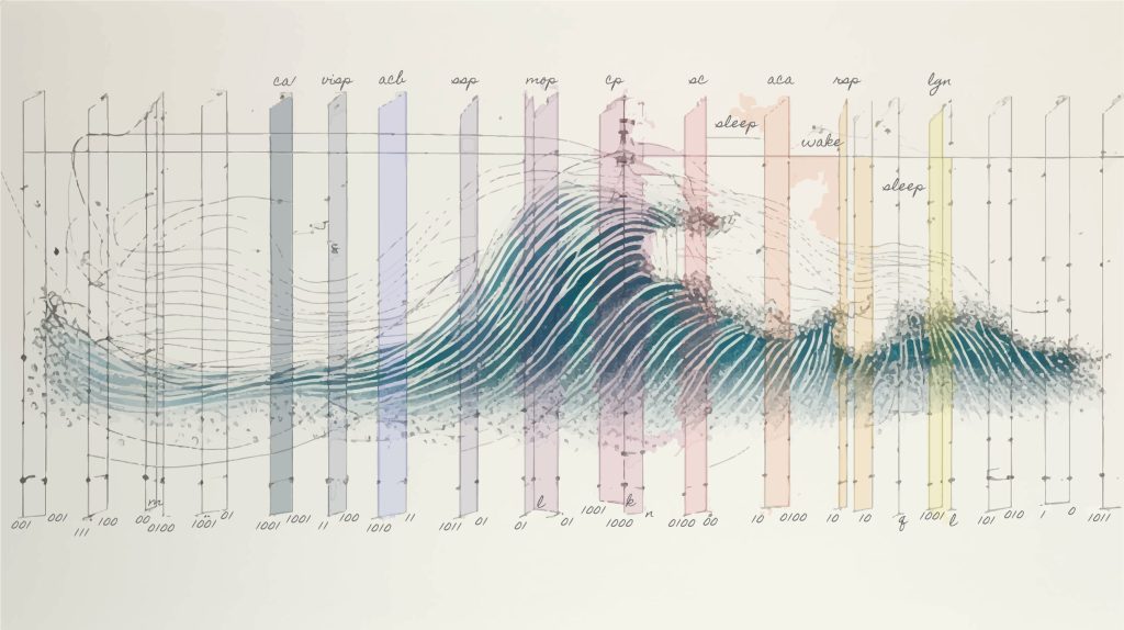
Hengen’s artistic interpretation of the different brainwave patterns that produce the basic states of sleep and wakefulness. Credit: Keith Hengen
Sleep and wakefulness. These are completely different states that define the boundaries of our daily lives. For years, scientists have been measuring the differences between these instinctive brain processes by observing brainwaves. Sleep is characterized by slow, prolonged waves measured in tenths of a second that travel throughout the organs.
For the first time, scientists have discovered that sleep can be detected by neural activity patterns that last just a few milliseconds – a thousandth of a second – revealing new ways to study and understand the fundamental brainwave patterns that govern consciousness. They have also shown that small areas of the brain can momentarily “flicker” awake while the rest of the brain remains asleep, and vice versa, changing from an awake to an asleep state.
These findings are New Research Published in the journal Nature Neuroscienceis a collaboration between the laboratories of Keith Hengen, assistant professor of biology at Washington University in St. Louis, and David Haussler, distinguished professor of biomolecular engineering at the University of California, Santa Cruz. The research was conducted by doctoral students David Parks (University of California, Santa Cruz) and Aidan Schneider (Washington University).
Over four years of research, Parks and Schneider trained neural networks to study patterns within vast amounts of EEG data, discovering patterns that occurred with unprecedented frequency and called into question long-held fundamental concepts about the neurological basis of sleep and wakefulness.
“With powerful tools and new computational methods, we have a lot to gain from questioning our most fundamental assumptions and rethinking the question, ‘what is a state?'” Hengen says. “Sleep and wakefulness are the biggest determinants of behavior, and everything else falls outside of that. So if we don’t understand what sleep and wakefulness actually are, we’re missing out.”
“For us as scientists, it was a surprise to find that different parts of the brain actually take a little nap while the other parts are awake. But many people may have already suspected that this phenomenon occurs in their spouses, so maybe the lack of gender bias is what’s surprising,” Hausler joked.
Understanding Sleep
Neuroscientists study the brain through recording its electrical signals. Brain activityIn this data, known as electrophysiology data, we see voltage waves that rise and fall at different paces, mixed with the spiking patterns of individual neurons.
The researchers worked with data from mice at the Hengen Institute in St. Louis, outfitting the freely moving animals with extremely lightweight headsets that recorded brain activity in 10 different brain regions for months at a time, tracking voltages from small populations of neurons with microsecond precision.
Feeding this amount of data created a petabyte (a million times gigabytes) of data. David Parks led the effort to feed this raw data into an artificial neural network that can find highly complex patterns, distinguish between sleep and awake data, and spot patterns that might be missed by human observation. Working with a shared academic computing infrastructure at the University of California, San Diego, the team was able to work with large volumes of data on the scale used by large companies like Google and Facebook.
Knowing that sleep is traditionally defined by slow-moving waves, Parks began feeding the neural network smaller and smaller chunks of data, asking it to predict whether the brain was asleep or awake.
The team found that the model could distinguish between sleep and wakefulness from just a few milliseconds of brain activity data. This was shocking to the team: it showed that the model couldn’t have relied on slow-moving waves to learn the difference between sleep and wakefulness. If you hear a thousandth of a second of a song, you can’t tell if it’s a slow rhythm. Similarly, a model can’t learn a rhythm that occurs over several seconds by looking at only a few randomly separated milliseconds of information.
“We’re looking at a level of detail that we’ve never seen before,” Hausler said. “Previously, the idea was that we wouldn’t find anything there, that all the relevant information would be in the lower frequency waves. This paper shows that if we ignore the traditional measurements and only look at the details of the high frequency measurements, at milliseconds, we have enough information to tell us whether the tissue is asleep or not. This shows that something is happening on a very fast scale, and gives us another hint as to what happens during sleep.”
Hengen, on the other hand, was convinced that Parks and Schneider’s findings were so at odds with basic concepts drilled into him from years of neuroscience education that he was sure they were missing something, and he challenged Parks to provide more and more evidence that the phenomenon might be real.
“This made me ask myself, ‘To what extent are my beliefs based on evidence, and what evidence do I need to overturn them?'” Hengen said. “It really felt like a game of cat and mouse, because I told David, [Parks] “Over and over again he would present me with evidence and try to get me to prove things, and he would come back and say, ‘Look at this.’ It was a really interesting process for me as a scientist to have my students tear down these towers, brick by brick, and then have to accept that.”
Local Patterns
artificial neural network Because machine learning is essentially a black box and doesn’t report what it’s learned, Parks began peeling back layers of temporal and spatial information to try to understand what patterns the model was learning.
Eventually, they got to the point where they were looking at chunks of brain data just one millisecond long, and the highest frequencies of brain voltage fluctuations.
“We took all the information that neuroscience has used over the last century to understand, define and analyze sleep and asked, ‘Can this model learn under these conditions?'” Parks says. “This allowed us to look at signals that we couldn’t understand before.”
Looking at this data, the researchers were able to determine that ultrafast patterns of activity among just a few neurons were the building blocks of sleep that their model detected. Importantly, such patterns could not be explained by traditional slow, widespread waves. The researchers hypothesized that the slow-moving waves might play a role in coordinating the faster, more localized patterns of activity, but ultimately concluded that the faster patterns were much closer to the essence of sleep.
If we compare the slow waves traditionally used to define sleep to thousands of people waving at a baseball stadium, these fast patterns are the conversations taking place between just a few people who decide to join the wave. The conversations are essential to the overall larger wave and are more directly related to the atmosphere in the stadium; the waves are a secondary result of that.
Observe the flicker
As they studied the hyperlocal patterns of activity further, the researchers began to notice another surprising phenomenon.
While looking at models that predict sleep and wakefulness, the researchers noticed what at first seemed like errors: for a split second, the model would detect that one area of the brain was awake, while the rest of the brain remained asleep. They observed the same thing during wakefulness: for a split second, one area would fall asleep while the rest of the brain remained awake. The researchers call these instances “flickering.”
“If you look at the time each of these neurons fires individually, [the neurons] “You’re going into a different state,” Schneider said, “and in some cases, these flickers can be localized to certain areas of the brain, and they can be even smaller than that.”
This prompted researchers to investigate what flicker means about sleep function and how it affects behavior during sleep and wakefulness.
“There’s a natural hypothesis there: Let’s say part of your brain falls asleep while you’re awake. Does that mean your behavior suddenly looks like you’re asleep? We’re starting to find that that’s often the case,” Schneider said.
By observing the behavior of mice, the researchers noticed that when parts of the brain flickered to sleep while the rest of the brain was awake, the mice would momentarily stop moving, as if they were drowsy. The sleep flickering (parts of the brain “waking up”) was reflected by the animals twitching while they slept.
Flicker is particularly surprising because it doesn’t follow the well-established rules that dictate the strict cycle in which the brain moves from wakefulness to non-REM sleep to REM sleep.
“We see all sorts of combinations — blinking from wakefulness to REM sleep, blinking from REM sleep to non-REM sleep — breaking the rules that we would expect based on 100 years of literature,” Hengen says. “We think it reveals a dissociation between the macrostates of sleep and wakefulness at the whole-animal level and the fast and local patterns that are the fundamental units of state in the brain.”
Impact
A deeper understanding of the patterns that occur at high frequencies, and the flickering between wakefulness and sleep, could help better study neurodevelopmental and neurodegenerative diseases that are associated with sleep dysregulation. Both Hausler’s and Hengen’s research groups are interested in understanding this connection further, and Hausler is interested in studying these phenomena further in brain organoid models, which are pieces of brain tissue grown on the bench.
“This could be a very sharp scalpel for solving questions about disease and disability,” Hengen said. sleep The higher the level of arousal, the more likely it is that appropriate clinical and disease-related questions will be addressed.”
At a fundamental level, this research helps to improve our understanding of the complexity of the brain’s many layers as an organ that determines behavior, emotions, and more.
For more information:
David F. Parks et al., “Nonoscillatory Millisecond Embedding of Brain States Provides Insights into Behavior.” Nature Neuroscience (2024). DOI: 10.1038/s41593-024-01715-2
Provided by
University of California, Santa Cruz
Quote: Scientists discover that small regions of the brain can take micronaps while the rest of the brain is awake, and vice versa (July 16, 2024) Retrieved July 17, 2024 from https://medicalxpress.com/news/2024-07-scientists-small-regions-brain-micro.html
This document is subject to copyright. It may not be reproduced without written permission, except for fair dealing for the purposes of personal study or research. The content is provided for informational purposes only.


